

Amblyopia, commonly known as “lazy eye,” is a vision development disorder in which one eye does not achieve normal visual acuity, often without noticeable symptoms. A person may function normally in daily life while the brain gradually favors the stronger eye, making the condition difficult to detect. It affects approximately 2–3% of the global population.
Although it typically begins in childhood, amblyopia can persist into adulthood if not treated early. While early detection offers the best outcomes, treatment may still provide benefits later in life depending on the individual case.
This guide explains what amblyopia is, how it develops, and the available treatment options for both children and adults.
What is amblyopia?
Amblyopia is a neurodevelopmental visual disorder characterized by reduced visual acuity in one eye (unilateral amblyopia) or, less commonly, both eyes (bilateral amblyopia), which cannot be fully corrected by optical means alone. Unlike refractive errors that can be resolved with glasses, amblyopia stems from abnormal visual development during a critical period of brain maturation, typically the first 7–8 years of life.
During normal visual development, the brain learns to process and integrate signals from both eyes simultaneously. In amblyopia, this process is disrupted: the brain begins to suppress or ignore the input from one eye, either because the image is blurred, misaligned, or blocked, leading to a functional "disconnection" between that eye and the visual cortex.
Key insight: Amblyopia is not a problem with the eye itself, but with how the brain learns to see. The eye is structurally normal in most cases; the deficit lies in neural processing.
The term "lazy eye" is widely used but can be misleading, implying a character flaw or voluntary disengagement. In reality, the eye is not lazy; it is undertrained. The brain has simply learned to favor the dominant eye, leaving the amblyopic eye's neural pathways underdeveloped.
Types of amblyopia
Ophthalmologists typically classify amblyopia into two main categories:
Functional amblyopia
This is the most common form, accounting for the vast majority of cases. It occurs when amblyopia develops in an otherwise healthy eye due to abnormal visual experience, without any underlying structural ocular pathology. Functional amblyopia includes:
- Strabismic amblyopia: caused by ocular misalignment (strabismus), where the brain suppresses input from the deviating eye to avoid double vision.
- Refractive amblyopia: caused by a significant uncorrected refractive error (usually high hyperopia or astigmatism) in one or both eyes. Anisometropic amblyopia, a subtype, occurs when a major difference in refractive error between the two eyes causes the brain to suppress the blurrier image.
- Meridional amblyopia: caused by uncorrected astigmatism, where certain visual orientations are selectively blurred.
Organic amblyopia
This form is caused by an identifiable structural abnormality that physically prevents normal visual input. Common causes include:
- Congenital or pediatric cataracts (opacity of the lens)
- Ptosis (drooping of the upper eyelid) obstructing the visual axis
- Corneal opacities or other media opacities
Organic amblyopia tends to be more severe and can be harder to treat, particularly if the underlying condition is not addressed surgically at an early stage.
Causes and risk factors
Any condition that interferes with clear, aligned, or symmetric visual input during early childhood can trigger amblyopia. The most common causes include:
Ocular misalignment (strabismus): Strabismus
When one eye turns inward (esotropia), outward (exotropia), upward, or downward, the brain receives two conflicting images. To avoid diplopia (double vision), the brain progressively suppresses the image from the misaligned eye. Strabismus is responsible for amblyopia in approximately 30–40% of cases.
Astigmatism and other refractive errors
High or asymmetric astigmatism , a condition where the cornea or lens has an irregular curvature, produces a consistently blurred retinal image, which the brain may learn to ignore. Similarly, high uncorrected hyperopia (farsightedness) or myopia (nearsightedness) in one eye can lead to deprivation of clear vision.
Stimulus deprivation
Physical obstruction of vision, from a cataract, ptosis, or other media opacity, prevents the developing visual system from receiving adequate stimulation. Deprivation amblyopia, though less common, is often the most severe form and requires urgent treatment.
Clinical note: A family history of strabismus, amblyopia, or significant refractive error significantly increases a child's risk and warrants earlier ophthalmological screening.
Recognizing the symptoms: Children vs. adults
Symptoms in children
One of the greatest diagnostic challenges in amblyopia is that young children, particularly those under 6 years old, rarely report visual problems. A child who has always seen the world through one dominant eye has no frame of reference for "normal" binocular vision. Parents and caregivers must therefore be vigilant for indirect signs:
- Squinting or closing one eye, especially in bright light or when trying to focus
- Head tilting or turning to favour one side
- Frequent eye rubbing or blinking of one eye
- Poor depth perception (difficulty judging distances, clumsiness)
- Complaints of headaches or eye fatigue, particularly after reading
- Apparent difficulty reading, copying from a board, or following moving objects
Because children adapt remarkably well to monocular vision, the condition can progress unnoticed for years without formal screening. This is why routine visual screening from the age of 6–12 months, and again at preschool entry, is strongly recommended by paediatric ophthalmological associations worldwide.
Symptoms in Adults
In adults who have not received treatment during childhood, amblyopia manifests primarily as reduced visual acuity that cannot be corrected to 20/20 with glasses or contact lenses. Depending on severity, this may significantly impact daily activities, including driving, reading, and professional performance.
Adults with amblyopia may also experience:
- Difficulty with tasks requiring fine visual discrimination
- Reduced contrast sensitivity and depth perception
- Increased visual fatigue and eye strain
Clinically, amblyopia is stratified into three levels of severity:
- Mild amblyopia: visual acuity of 0.5 to 0.8 (6/12 to 6/7.5)
- Moderate amblyopia: visual acuity of 0.2 to 0.5 (6/30 to 6/12)
- Severe/profound amblyopia: visual acuity of 0.1 or below (6/60 or worse)
Diagnosis: Screening and evaluation methods
Early and accurate diagnosis is the cornerstone of effective amblyopia management. The diagnostic approach depends on the patient's age and ability to cooperate:
In infants and pre-verbal children
- Photoscreening: a camera-based technique that assesses the red reflex from each eye simultaneously, detecting risk factors such as anisometropia, media opacities, and large refractive errors
- Pupillary reflex testing: assesses the symmetry of the pupillary light response to identify asymmetry suggesting a sensory deficit
- Preferential looking tests: measure visual acuity indirectly by observing the child's tendency to look at patterned stimuli rather than plain stimuli
In older children and adults
- Best-corrected visual acuity (BCVA) measurement: the primary diagnostic criterion; a 2-line or greater difference between eyes on a Snellen chart is a key diagnostic indicator
- Crowding effect testing: amblyopic eyes often perform disproportionately poorly when letters are presented closely together (the crowding phenomenon), a hallmark of amblyopia
- Cover-uncover test and alternate cover test: to detect strabismus
- Cycloplegic refraction: performed under mydriatic drops to measure the full refractive error without accommodation
Important: Amblyopia is a diagnosis of exclusion, structural eye disease must be ruled out before attributing reduced visual acuity to amblyopia alone.
Treatment options: From patching to cutting-edge therapies
The treatment of amblyopia is guided by the underlying cause, the patient's age, and the severity of visual loss. The central principle is the same across all approaches: stimulate the amblyopic eye by reducing the dominance of the stronger eye.
Refractive correction
The first step in treating any form of amblyopia is to optimise visual input by correcting the underlying refractive error with spectacles or contact lenses. In mild to moderate cases, optical correction alone, maintained consistently over several months, can significantly improve visual acuity, as the brain receives a clearer image from the amblyopic eye and begins to re-engage it.
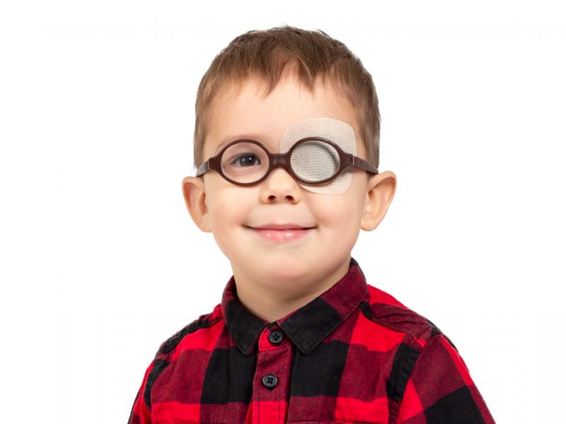
Occlusion therapy (eye patching)
Patching the dominant (non-amblyopic) eye remains the gold standard of amblyopia treatment. By covering the stronger eye for a prescribed number of hours per day, the child is forced to rely on the amblyopic eye, driving neuroplastic changes in the visual cortex. Treatment duration ranges from weeks to months, and compliance, a major clinical challenge, is critical to outcomes.
- Typical patching duration: 2–6 hours per day for moderate amblyopia
- Patching should be combined with near visual activities (drawing, reading, puzzles) to maximise cortical stimulation
Atropine penalization
When patching is poorly tolerated or impractical, the application of atropine eye drops to the sound eye provides an alternative. Atropine temporarily blurs near vision in the dominant eye by dilating the pupil and paralyzing accommodation, thereby forcing the amblyopic eye to do the work of seeing at close range. Studies have shown atropine penalization to be comparably effective to patching in moderate amblyopia.
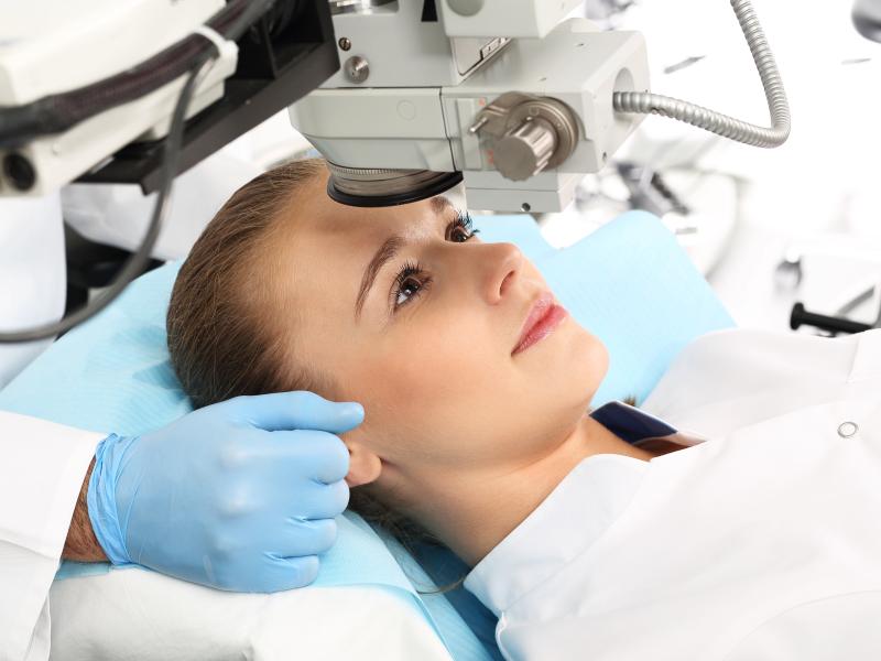
Surgical intervention
When amblyopia is secondary to strabismus, surgical correction of the ocular misalignment is often recommended to restore binocular alignment and create the conditions for effective patching therapy. For organic amblyopia caused by cataracts or ptosis, prompt surgical removal of the obstruction is essential, ideally within the first weeks of life, to prevent irreversible deprivation amblyopia.
Dichoptic training and digital therapies (emerging)
An exciting area of current research involves dichoptic training: computer-based or game-based visual therapy that presents different images to each eye simultaneously, training the brain to use both eyes together. Studies suggest this approach can improve visual acuity and binocularity, particularly as an adjunct to conventional therapy.
Treatment in adults
While the critical period for amblyopia treatment traditionally ends around age 7–8, accumulating evidence suggests that neuroplasticity persists into adulthood to a greater degree than previously thought. Adult amblyopia treatment has demonstrated modest but clinically meaningful improvements in visual acuity, particularly through perceptual learning programmes, dichoptic therapy, and, in selected cases, patching. Treatment expectations must be realistic, but the field is evolving rapidly.
Treatment is most effective when initiated early during the critical period of visual development (before approximately 7 years of age), with reported response rates often exceeding 70–80% in young children. Outcomes tend to become more variable with increasing age; nevertheless, meaningful improvement remains possible even in older children and adults (Yeritsyan et al., Cureus, 2024, PubMed PMID: 38650802)
Prevention and the importance of regular eye screenings
Amblyopia cannot always be prevented, some causes, like congenital cataracts, are unavoidable. However, the progression to significant visual loss is almost always preventable through timely detection and treatment.
Key preventive measures include:
- Neonatal and infant eye examinations: checking for red reflex, media clarity, and early signs of strabismus from birth
- Preschool vision screening (ages 3–5): ideally using standardised chart-based testing or photoscreening
- School-entry vision assessments
- Regular follow-up for children with a family history of strabismus, amblyopia, or refractive error
- Early and consistent correction of refractive errors with appropriate glasses
Parents are encouraged to observe their children's visual behavior and seek specialist assessment without delay if any of the warning signs described in this article are noted.

"Medical journalist specializing in science communication, I put my expertise at the service of clear and accessible information. For Turquie Santé, I create content based on up-to-date medical data, in collaboration with specialists from partner clinics. My commitment is to provide reliable, transparent information that complies with international medical standards."
Need a personalized medical opinion?
Our partner doctors reply online within 24h, free of charge.