


Vision disorders affect hundreds of millions of people worldwide, with more than 2.2 billion individuals living with some form of visual impairment according to the World Health Organization. In response to this major global health challenge, modern ophthalmology has made significant progress, particularly through corneal transplantation and amniotic membrane transplantation (AMT). These two surgical techniques now provide effective solutions for restoring or preserving sight.
In this guide, we explain in clear, accessible terms how these procedures work, when they are indicated, and what clinical outcomes patients can expect. We also explore why Turkey has emerged as a leading destination for high-quality, affordable ophthalmic care.
The role of the cornea in vision
The cornea is the transparent, dome-shaped surface that covers the front of the eye. It is responsible for 65 to 75% of the eye's total refractive power, making it the primary focusing structure of the visual system. When healthy, it bends incoming light precisely onto the retina for sharp, clear vision.
Various diseases can compromise corneal clarity or structure: keratoconus (progressive thinning), Fuchs' endothelial dystrophy (dysfunction of the inner cell layer), infectious scarring (herpes simplex, bacteria), trauma, or chemical burns. When spectacles, rigid contact lenses, or medical therapy are insufficient, surgical intervention is considered.
Two major surgical strategies exist: keratoplasty (corneal transplant) and amniotic membrane transplantation (AMT). Their indications, mechanisms of action, and risk profiles differ fundamentally.
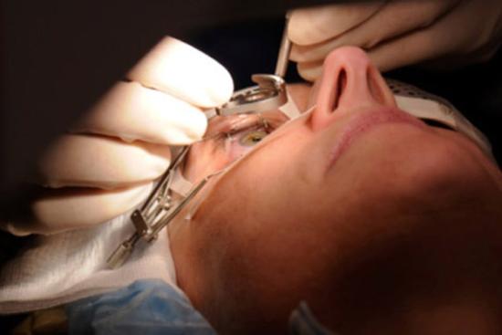
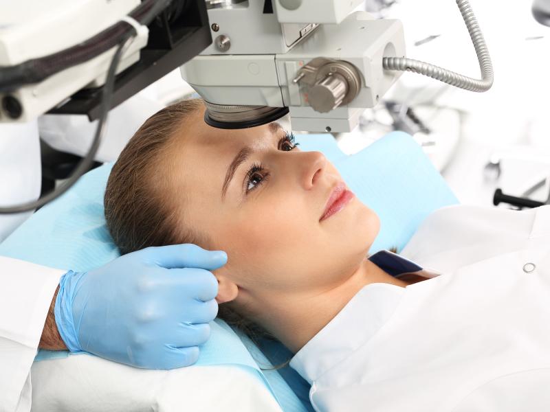
Corneal transplant procedures (keratoplasty)
Keratoplasty replaces all or part of a diseased cornea with healthy donor tissue. Techniques have evolved towards increasingly selective, layer-specific grafts, which reduces rejection risk and improves visual outcomes.
Penetrating keratoplasty (PK), full-thickness graft
PK is the historical technique: the surgeon removes the full central thickness of the cornea and replaces it with a sutured full-thickness graft. Although well-established, it carries a longer recovery (12–18 months), a higher rejection risk, and suture-induced irregular astigmatism.
A 2019 meta-analysis published in Ophthalmology reported a 5-year graft survival rate of 72% for PK, compared to over 85% for modern lamellar techniques. PK remains indicated when multiple corneal layers are simultaneously affected.
Lamellar keratoplasty, targeted partial grafts
Lamellar techniques replace only the diseased corneal layer while preserving healthy layers. This approach significantly reduces rejection risk, the endothelium–Descemet complex is the primary trigger of immune response, and shortens recovery.
DSAEK, Descemet's Stripping Automated Endothelial Keratoplasty
DSAEK replaces the endothelium and a thin posterior stromal layer with a graft approximately 100–150 µm thick. Tissue is inserted through a small incision (3–4 mm) and fixed with an air bubble. Stable visual recovery is typically achieved in 3 to 6 months. A study published in Cornea (2020) reported a mean visual acuity gain of +0.3 LogMAR at 12 months.
DMEK, Descemet Membrane Endothelial Keratoplasty
DMEK is currently considered the gold standard for isolated endothelial conditions (Fuchs' dystrophy, bullous keratopathy). It transplants only the Descemet membrane and endothelium, a tissue layer less than 15 µm thick.
Data published in JAMA Ophthalmology (Price et al., 2021) show that 90% of DMEK patients achieve visual acuity ≥ 20/25 at 1 year, with a rejection rate below 1%. Visual recovery is faster than with DSAEK: functional acuity is often reached within 4 to 8 weeks.
DALK, Deep Anterior Lamellar Keratoplasty
DALK replaces the anterior stroma while preserving the patient's own endothelium. It is particularly indicated in advanced keratoconus and stromal scarring. By sparing the patient's endothelium, it virtually eliminates the risk of endothelial rejection, the leading cause of long-term graft failure.
Patient Story, Emilia, 34, Marseille “I was diagnosed with advanced bilateral keratoconus, and my ophthalmologist in France recommended a full-thickness corneal transplant with an 18-month waiting list. Through Turquie Santé, I underwent DALK surgery in Istanbul within just six weeks, with seamless coordination before and after my return. Six months later, I can read without lenses for the first time in ten years.”
Patient testimonial, 2025, name changed with consent. Results may vary depending on individual medical conditions.
Main indications for corneal transplant
- Keratoconus (progressive corneal thinning)
- Fuchs' endothelial dystrophy
- Post-surgical bullous keratopathy
- Corneal scarring (herpes, trauma, deep burns)
- Corneal oedema refractory to medical treatment
- Failed previous graft
Amniotic membrane transplantation (AMT)
The amniotic membrane is the innermost layer of the human placenta. After rigorous screening (infection testing, processing, irradiation or cryopreservation), it is used as a surgical biomaterial on the ocular surface.
AMT is not a corneal transplant: it does not replace the corneal structure, but creates a biologically favourable environment for ocular surface repair. It is often used as a complement to, or preparation for, corneal transplantation.
Documented biological properties of amniotic membrane
- Anti-inflammatory action: suppression of pro-inflammatory cytokines (IL-1, TNF-α), beneficial in chemical burns and severe inflammation.
- Re-epithelialization support: the membrane acts as a substrate for epithelial cell migration; in vitro studies published in Investigative Ophthalmology & Visual Science show healing acceleration of 40 to 60% compared to conventional care.
- Anti-scarring activity: inhibition of myofibroblasts, reducing opacifying fibrous tissue formation.
- Analgesic effect: reduction of post-operative pain, sometimes within the first 48 hours.
- Antimicrobial properties: presence of peptides (defensins, elafin) active against certain bacteria and fungi.
Because amniotic membrane is acellular after processing, the risk of immunological rejection is very low, a fundamental distinction from corneal grafts, where long-term local immunosuppression is essential.
Main indications for AMT
- Chemical or thermal eye burns (urgent indication)
- Persistent epithelial defects resistant to conventional treatment
- Refractory corneal ulcers
- Limbal stem cell deficiency
- Severe dry eye disease (DEWS II stage III–IV)
- Recurrent pterygium
- Symblepharon (conjunctival adhesions)
- Ocular surface preparation before corneal transplant
Comparison table: corneal transplant vs AMT
The table below summarises the main clinical differences between the two approaches to help physicians and patients identify the most appropriate strategy.
| Criterion | Corneal transplant (keratoplasty) | Amniotic membrane graft (AMT) |
| Primary goal | Replace structurally failing corneal tissue | Promote ocular surface healing |
| Tissue source | Deceased human donor cornea (tissue bank) | Processed placental membrane (cryopreserved) |
| Invasiveness | Moderate to high (technique-dependent) | Low (suture or biological glue) |
| Anaesthesia | General or peribulbar block | Topical or local |
| Recovery | 4–8 weeks (DMEK) to 12–18 months (PK) | Days to a few weeks |
| Rejection risk | Present, long-term topical steroids required | Very low (acellular tissue after processing) |
| Typical indications | Keratoconus, Fuchs' dystrophy, stromal scarring | Burns, ulcers, severe dry eye, limbal deficiency |
| Combined use | Yes, AMT often prepares the surface before keratoplasty | Yes |
Overall, the choice between these two techniques depends on the severity and nature of corneal damage, with amniotic membrane transplantation often used as an initial or supportive step, while corneal transplantation is reserved for cases requiring structural replacement of the cornea.
Turkey: a reference destination for eye surgery
Recent data indicate that Turkey receives well over 1 million international patients annually, with estimates ranging from approximately 1.4 million to nearly 2 million health tourists in recent years, according to official communications from Turkish authorities (Republic of Türkiye Directorate of Communications, 2024; Turkish Ministry of Health).
- Surgical expertise and international accreditations : Turquie Santé's partner ophthalmology centres are JCI (Joint Commission International) accredited, a leading international standard for quality and patient safety. Surgeons are trained in Europe and North America and contribute to peer-reviewed publications.
- State-of-the-art equipment : Partner hospitals use advanced ocular microsurgery technologies such as femtosecond lasers, intraoperative OCT, and high-definition specular microscopy. Certified tissue banks ensure traceability and quality control of donor corneas.
- Research and innovation: University hospitals in Istanbul, Ankara and Izmir are involved in research on corneal tissue engineering, bioartificial keratoprostheses, and amniotic membrane technologies. Ankara University Eye Hospital participates in European clinical trials on limbal stem cell deficiency.
- Cost and accessibility: Procedures such as DMEK or DSAEK typically range from €2,500 to €5,000 all-inclusive in Turkey, compared to €8,000–€15,000+ in Western Europe, largely due to lower operational costs rather than differences in medical quality or standards.
Important clinical nuances
Every surgical decision must be individualised. The data presented in this article reflect average results from the scientific literature; individual outcomes depend on many factors: disease stage, ocular surface condition, age, comorbidities, and surgeon experience.
- AMT is not a definitive treatment for structural disease: in advanced keratoconus or endothelial dystrophy, it can prepare the surface but does not replace the need for keratoplasty.
- Rejection risk, though low with DMEK, is not zero: regular ophthalmological monitoring and long-term topical corticosteroid treatment remain essential.
- DMEK is technically demanding: the best outcomes are achieved in high-volume surgical centres, which underscores the importance of centre and surgeon selection.
- Results may vary with aetiology: corneal oedema from trauma responds differently from genetic dystrophy. Thorough preoperative assessment is essential.

"As I am passionate about science, I studied optometry which I eventually gave up. In the midst of it all, I fell in love with the editorial staff.I think that the best form of generosity is to share your knowledge after consulting a number of encyclopedias. "
Need a personalized medical opinion?
Our partner doctors reply online within 24h, free of charge.