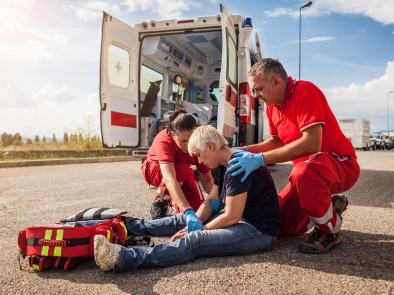
Stroke is the second leading cause of death worldwide and the single largest cause of acquired adult disability. Yet not all strokes are the same, and treating one type like the other can be catastrophic. The drug that dissolves a clot in an ischemic stroke can trigger fatal bleeding in a hemorrhagic one. This is why distinguishing between the two is not merely an academic exercise: it is a time-critical clinical decision made within minutes of a patient's arrival in the emergency room.
Every year, 15 million people suffer a stroke globally. Of these, 5 million die and another 5 million are left permanently disabled, according to the World Stroke Organization. The good news: 80% of strokes are preventable. The even better news: when treated promptly and correctly, outcomes have improved dramatically over the past decade thanks to thrombectomy, advanced neuroimaging, and organized stroke units.
This medically reviewed guide explains the biology, warning signs, diagnosis, and treatment of both stroke types, and outlines what high-quality, international-standard care looks like today.
The core difference: Two mechanisms, two emergencies
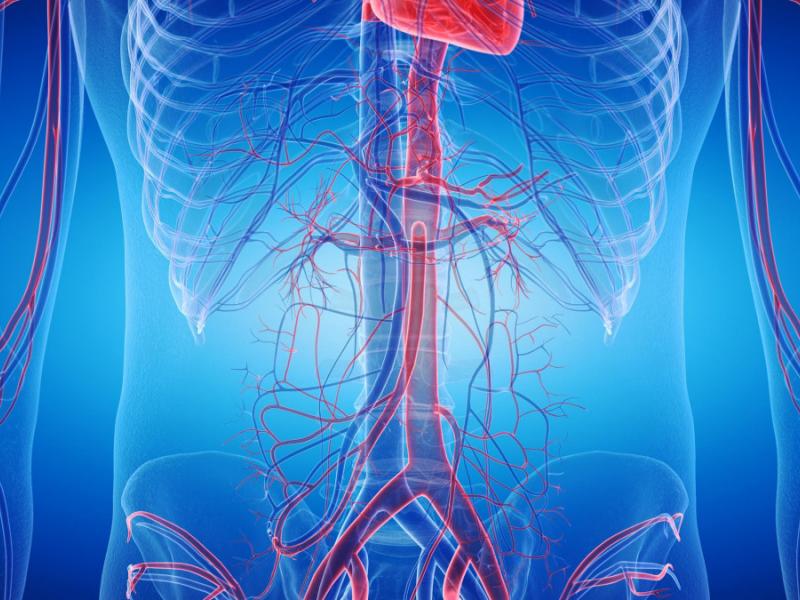
At its most fundamental, an ischemic stroke happens when a blood vessel supplying the brain is blocked. A hemorrhagic stroke happens when a blood vessel ruptures. In ischemic stroke, neurons starve. In a hemorrhagic stroke, they drown. Both are lethal. Both are time-sensitive. Both require immediate responses, but they are entirely different.
| Criterion | Hemorrhagic stroke | Ischemic stroke |
| Mechanism | Burst blood vessel → brain bleed | Blocked blood vessel → oxygen deprivation |
| Frequency | ~20% of all strokes | ~80% of all strokes |
| Onset | Sudden - 'thunderclap' headache | Gradual or sudden - motor deficit |
| Key imaging | Non-contrast CT (hyperdensity = blood) | CT + MRI (may be normal in the first hour) |
| Urgent treatment | Blood pressure control + surgery if needed | IV thrombolysis (4.5 h) or thrombectomy |
| Top risk factor | Uncontrolled hypertension | Atrial fibrillation + atherosclerosis |
| 30-day mortality | 40 – 50% | 15 – 20% |
| Recovery potential | Slower, but the long-term outcome can be good | Better if treated within the therapeutic window |
The table above captures the essential clinical asymmetry: ischemic strokes are far more common but more treatable within a tight time window; hemorrhagic strokes are rarer but carry a much higher early mortality, and their treatment is largely about limiting damage rather than reversing it.
The biology behind each type
Although ischemic and hemorrhagic strokes can produce similar symptoms, the biological mechanisms behind them are fundamentally different.
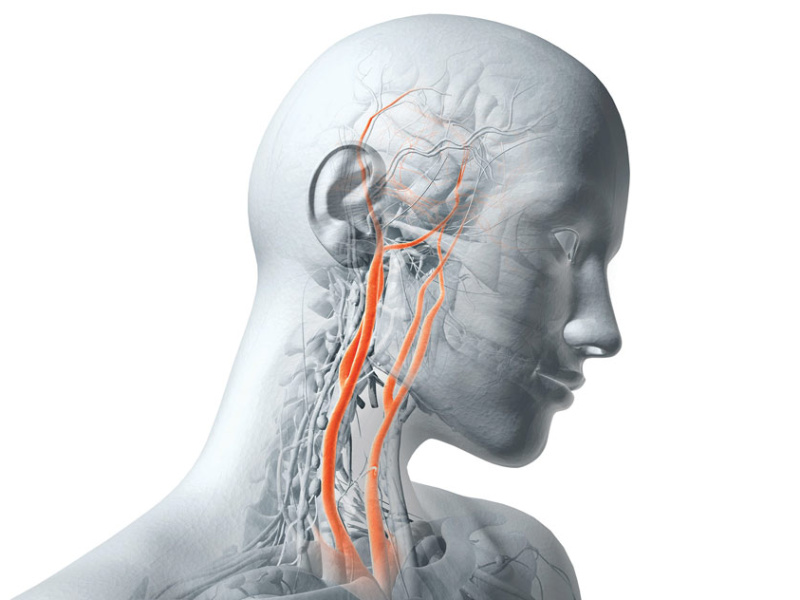
Ischemic stroke: When blood flow stops
In an ischemic stroke, a blood clot blocks an artery supplying the brain. The clot may form locally on an atherosclerotic plaque (thrombosis) or travel from elsewhere in the body (embolism), most often from the heart during atrial fibrillation. Within minutes, the central area deprived of blood supply undergoes irreversible necrosis.
Surrounding this core, however, lies the ischemic penumbra, a region of stunned but still viable tissue that can be rescued if circulation is restored quickly enough. This biological reality explains the extreme urgency of stroke treatment: the primary goal is to save the penumbra before it becomes permanently damaged.
Two main reperfusion strategies are available:
- Intravenous thrombolysis (alteplase / rt-PA), which chemically dissolves the clot, is effective when administered within 4.5 hours after symptom onset.
- Mechanical thrombectomy, a catheter-based procedure that retrieves the clot under fluoroscopic guidance, can extend the therapeutic window to up to 24 hours in selected patients.
Mechanical thrombectomy is considered one of the most important advances in stroke medicine since the introduction of thrombolytic therapy in 1996, dramatically improving outcomes in patients with large-vessel occlusions.
Hemorrhagic stroke: When the vessel fails
Hemorrhagic stroke occurs when a blood vessel ruptures, allowing blood to escape into or around the brain. Two main subtypes exist:
- Intracerebral hemorrhage (ICH), in which bleeding occurs directly within brain tissue
- Subarachnoid hemorrhage (SAH), in which blood floods the space between the brain and its surrounding membrane.
In both cases, the accumulating blood forms a hematoma that exerts increasing pressure on nearby brain structures. This pressure triggers a cascade of secondary injuries, including brain swelling (edema), rising intracranial pressure, and, in severe cases, brain herniation, a life-threatening condition.
The most common underlying cause of ICH is chronic, poorly controlled hypertension, which weakens the walls of small perforating arteries over time until they rupture. Cerebral aneurysms (the classic cause of SAH), arteriovenous malformations (AVMs), and certain medications, particularly anticoagulants, account for most of the remaining cases.
Recognising a stroke: Shared signs and key differentiators
Both types of stroke share the core FAST warning signs. However, certain symptoms are strong clinical pointers toward one type rather than the other. That said, and this cannot be overstated, no clinician can reliably distinguish hemorrhagic from ischemic stroke on symptoms alone. Only brain imaging can.
Warning signs common to both - FAST
The following symptoms should always be treated as a medical emergency:
- Sudden numbness or weakness of the face, arm, or leg, especially on one side of the body.
- Sudden confusion, difficulty speaking or understanding speech.
- Sudden vision loss in one or both eyes, or double vision.
- Sudden severe dizziness, loss of balance, or coordination.
- Sudden onset of severe headache with no known cause.
Symptoms more suggestive of a hemorrhagic stroke
Certain clinical features may raise suspicion of bleeding inside the brain:
- Thunderclap headache: a sudden, explosive headache reaching maximum intensity within seconds. Often described as “the worst headache of my life,” it is a classic sign of subarachnoid hemorrhage and should always be treated as an emergency until proven otherwise.
- Vomiting and neck stiffness, which may indicate irritation of the meninges caused by blood in the subarachnoid space.
- Rapid loss of consciousness, as an expanding hematoma quickly compresses critical brain structures.
- Severely elevated blood pressure, with systolic values frequently exceeding 180 mmHg at presentation.
Symptoms more suggestive of ischemic stroke
Some neurological patterns are more typical of an arterial blockage:
- Pure motor or sensory deficit, such as hemiplegia or hemisensory loss, developing over minutes, often without severe headache.
- Transient monocular blindness (amaurosis fugax): classically described as a “curtain descending over one eye.” This symptom often represents a transient ischemic attack (TIA) and may precede a major ischemic stroke.
- Isolated aphasia, a sudden inability to speak or understand language, typically resulting from ischemia in the left middle cerebral artery territory.
Critical clinical rule
Never administer anticoagulants or thrombolytics before brain imaging confirms the stroke type.
Giving thrombolytic therapy to a patient with hemorrhagic stroke can dramatically worsen bleeding and significantly increase mortality. For this reason, urgent CT imaging is one of the first steps in emergency stroke evaluation.
This decision, determining whether a stroke is ischemic or hemorrhagic, is among the most critical in emergency medicine, and it cannot be made on clinical signs alone.
Diagnosis: Speed and imaging are everything
The diagnostic protocol for acute stroke is designed around one objective: rule in or rule out hemorrhage as fast as possible. From door to CT should be under 25 minutes in any well-organised stroke centre.
Non-contrast CT scan: The gateway test
Non-contrast CT (NCCT) is the first-line imaging modality in acute stroke. It detects intracerebral hemorrhage with near-perfect sensitivity. Fresh blood appears as a hyperdense (white) region on CT within minutes of the bleed. An ischemic infarct, however, may be entirely invisible on NCCT within the first few hours, which is why a 'normal CT' does not exclude stroke and should never delay treatment when clinical suspicion is high.
MRI with diffusion weighting (DWI)
DWI-MRI detects ischemic core tissue within minutes of onset, far earlier than conventional MRI sequences. It is the most sensitive tool for small, deep, or posterior fossa infarcts that CT often misses. When combined with perfusion imaging (PWI), it maps the ischemic penumbra and helps determine whether a patient is likely to benefit from late-window thrombectomy beyond the standard 6-hour threshold.
CT angiography and digital subtraction angiography
CTA (CT angiography) is performed immediately after the initial NCCT to identify large vessel occlusion (LVO) in suspected ischemic stroke. This finding predicts a poor outcome with medical treatment alone and mandates consideration of thrombectomy. In hemorrhagic stroke, CTA identifies aneurysms, AVMs, and the 'spot sign' (active contrast extravasation predicting hematoma expansion). Digital subtraction angiography (DSA) remains the gold standard for preprocedural vascular assessment before aneurysm coiling or surgical clipping.
Risk factors: What you can and cannot change
Eight modifiable risk factors account for more than 90% of stroke risk worldwide (O'Donnell et al., Lancet 2010; INTERSTROKE study). The implication is clear: stroke prevention is largely within our control.
| Risk factor | Stroke type | Modifiable? | Clinical action |
| Uncontrolled hypertension | Both | Modifiable | Largest single risk factor |
| Atrial fibrillation | Ischemic | Modifiable | Anticoagulation therapy |
| Smoking | Both | Modifiable | Risk halves within 2 years |
| Type 2 diabetes | Both | Modifiable | HbA1c < 7% target |
| Dyslipidemia | Ischemic | Modifiable | Statins, diet |
| Excessive alcohol | Hemorrhagic | Modifiable | < 1 drink/day |
| Unsupervised anticoagulants | Hemorrhagic | Manageable | Regular INR monitoring |
| Age > 55 | Both | Fixed | More frequent screening |
| Family history of stroke | Both | Fixed | Early risk assessment |
The single most impactful preventive intervention remains blood pressure control. Keeping systolic BP below 130 mmHg reduces hemorrhagic stroke risk by approximately 40% and ischemic stroke risk by 25%. No drug, supplement, or lifestyle intervention comes close in terms of population-level benefit.
Treatment: Opposing strategies for two different crises
Treating ischemic stroke
The cornerstone of acute ischemic stroke treatment is reperfusion, restoring blood flow to the ischemic territory as quickly as possible.
- IV alteplase (0.9 mg/kg, max 90 mg) remains the standard thrombolytic when administered within 4.5 hours.
- Tenecteplase (TNK), a more convenient single-bolus alternative, is increasingly adopted in major stroke centres.
- Mechanical thrombectomy, endovascular retrieval of the offending clot using stent-retrievers or aspiration catheters under fluoroscopic guidance, is now indicated for large vessel occlusions up to 24 hours after onset in patients with salvageable penumbra on imaging. Its results are transformative: a 50% relative reduction in severe disability when performed promptly.
Secondary prevention begins immediately after stabilisation: antiplatelet therapy (aspirin ± clopidogrel for non-cardioembolic strokes), anticoagulation for atrial fibrillation (DOACs), statin therapy, and strict vascular risk factor management.
Treating hemorrhagic stroke
Management of hemorrhagic stroke focuses on hematoma containment and control of intracranial pressure. For mild-to-moderate bleeds, blood pressure reduction to <140 mmHg within the first hour is standard. Anticoagulation reversal is urgent when indicated (e.g., with vitamin K, prothrombin complex concentrate, or idarucizumab for dabigatran).
Surgical intervention, such as open craniotomy with clot evacuation or minimally invasive endoscopic surgery, is considered for large, accessible hematomas (>30 mL) that are causing mass effect.
The mainstay of treatment for subarachnoid hemorrhage from a ruptured aneurysm is aneurysm occlusion, either by :
- Endovascular coiling (the preferred method in most centers).
- Microsurgical clipping, performed during open neurosurgery
These procedures are usually carried out within 24 to 72 hours after the hemorrhage to minimize the risk of recurrent bleeding.
Cerebral vasospasm, a major cause of delayed neurological deterioration in SAH, is managed with nimodipine, induced hypertension, and endovascular balloon angioplasty.
Neurorehabilitation: The third pillar
Rehabilitation should begin within 24–48 hours of stroke stabilisation and should be as evidence-based as acute-phase treatments. Neuroplasticity, the brain's remarkable capacity to reorganise its neural circuits, is greatest in the first weeks and months after stroke.
A comprehensive programme includes physiotherapy for motor recovery, speech and language therapy, occupational therapy for functional independence, and neuropsychological support for the cognitive and emotional sequelae that affect up to 30% of survivors.
Stroke treatment in Turkey: Both types, one destination
Both ischemic and hemorrhagic strokes require highly specialized medical management, advanced imaging, and rapid multidisciplinary intervention. Over the past decade, Turkey has developed a strong reputation in neurovascular medicine, combining internationally trained specialists, modern hospital infrastructure, and significantly lower treatment costs.
Many leading hospitals operate comprehensive stroke centres equipped with advanced neuroimaging, interventional neuroradiology suites, and dedicated stroke units. Treatment costs are typically 40–60% lower than in Western Europe or North America, while maintaining high clinical standards.
Our JCI-accredited partner hospitals in Istanbul operate 24/7 Stroke Units staffed by experienced vascular neurologists, interventional neuroradiologists, and neurosurgeons. These multidisciplinary teams manage the full spectrum of stroke care, from diagnosis and acute treatment to long-term prevention and rehabilitation.
Patients may seek specialised care for:
- Post-stroke follow-up and neurological evaluation.
- Secondary stroke prevention and vascular risk assessment.
- Endovascular or surgical treatment of cerebral aneurysms or arteriovenous malformations (AVMs).
- Advanced neurorehabilitation programmes.
From the first medical inquiry to post-treatment follow-up, our international patient coordinators assist patients at every step. Fluent in English, French, and Arabic, they help organise medical evaluations, hospital appointments, and personalised care pathways to ensure a smooth and reassuring treatment experience.

"Medical journalist specializing in science communication, I put my expertise at the service of clear and accessible information. For Turquie Santé, I create content based on up-to-date medical data, in collaboration with specialists from partner clinics. My commitment is to provide reliable, transparent information that complies with international medical standards."
Need a personalized medical opinion?
Our partner doctors reply online within 24h, free of charge.

"An eminent Professor of cardiovascular surgery, Dr. Necmettin Çolak leads cutting-edge cardiac interventions in Ankara. Specialized in vascular pathologies and open-heart surgeries, he ensures our heart health articles reflect the latest clinical guidelines. His validation provides peace of mind for our readers."


Among our clinics