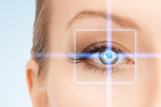


Ocular nystagmus is a condition that causes involuntary eye movements, making it difficult to keep the eyes focused on a fixed object. Although many people are unfamiliar with it, nystagmus affects around 1 in 1,000 individuals worldwide and can have a significant impact on daily life and vision quality.
Rather than being a disease itself, nystagmus is usually a sign of an underlying neurological, vestibular, or eye-related disorder. It may appear at birth or develop later in life, with symptoms ranging from mild visual disturbances to more noticeable vision problems.
In this article, we explore the causes, symptoms, diagnosis, and treatment options for ocular nystagmus, including recent advances in medical care and emerging therapies.
What is ocular nystagmus?
Ocular nystagmus is defined as a repetitive, involuntary, rhythmic oscillation of the eyes. These movements can occur in various directions, horizontal (most common), vertical, torsional (rotary), or a combination of these, and are generally beyond the individual's voluntary control.
Three characteristics define nystagmus:
- Oscillatory: movements occur side to side, up and down, or in a circular pattern
- Rhythmic: movements follow a regular, repetitive pattern
- Involuntary: the person is aware of the movements but cannot consciously stop them
Unlike a brief, momentary eye twitch, nystagmus is sustained and consistent. In many cases, individuals develop a "null point", a specific head position in which the eye movements are minimized and vision is at its clearest.
Symptoms of ocular nystagmus
Recognizing the symptoms early is key to timely diagnosis and management. While the involuntary eye movement is the defining feature, nystagmus presents with a broader constellation of signs that vary depending on the underlying cause and severity.
Visual symptoms
- Reduced visual acuity: Most people with nystagmus experience some degree of blurred or reduced vision, particularly at distance.
- Oscillopsia: A subjective sensation that the visual environment is moving or oscillating. This is more commonly reported in acquired nystagmus than in the congenital form.
- Photosensitivity: Sensitivity to bright light, especially in nystagmus associated with albinism.
- Difficulty with depth perception: Challenges judging distances, which can affect activities like driving or playing sports.
Physical and functional symptoms
- Abnormal head positioning: Many individuals unconsciously tilt or turn their head to find the null point where vision is most stable.
- Head nodding: Some children with congenital nystagmus develop rhythmic head nodding in compensation.
- Balance difficulties: Particularly in cases linked to vestibular dysfunction.
- Eye fatigue and headaches: Sustained effort to stabilize gaze can cause significant ocular and frontal discomfort.
Associated symptoms (Depending on cause)
- Dizziness or vertigo (vestibular nystagmus)
- Tinnitus or hearing loss (Ménière's disease, labyrinthitis)
- Neurological symptoms such as double vision, weakness, or coordination problems (central causes)
- Light sensitivity and nystagmus in very bright or very dim conditions (albinism-related)
Important: If nystagmus appears suddenly in an adult with no prior eye history, it should be treated as a medical emergency and evaluated immediately, it may indicate a stroke, brain hemorrhage, or acute neurological event.
Causes of ocular nystagmus
The underlying causes of nystagmus are diverse. In essence, nystagmus arises when the brain areas responsible for controlling smooth eye movements, primarily the cerebellum, brainstem, and vestibular system, receive disrupted or abnormal signals.
Congenital and hereditary causes
These forms usually appear during infancy or early childhood.
- Genetic mutations affecting the visual pathway (e.g., PAX6 gene mutations)
- Albinism (oculocutaneous or ocular), in which abnormal optic nerve decussation leads to nystagmus in nearly all affected individuals
- Congenital motor nystagmus (idiopathic infantile nystagmus), typically appearing within the first 6 months of life
Ocular pathologies
Certain eye conditions can interfere with normal visual development and trigger nystagmus.
- Congenital cataracts, when present in early infancy, they deprive the visual system of the stimulation needed for normal development
- Strabismus (misalignment of the eyes)
- Amblyopia (lazy eye)
- Optic nerve hypoplasia or degeneration
- High uncorrected refractive errors
Neurological causes
Nystagmus may also result from disorders affecting the brain or nervous system.
- Multiple sclerosis (MS): Nystagmus is reported in up to 70% of MS patients at some point in their disease course
- Brain tumors (particularly those affecting the cerebellum or brainstem)
- Stroke or transient ischemic attack (TIA)
- Head trauma
- Arnold-Chiari malformation
- Wernicke's encephalopathy (thiamine deficiency)
Vestibular causes
The vestibular system helps maintain balance and eye stability. Inner ear disorders can therefore lead to nystagmus.
- Benign paroxysmal positional vertigo (BPPV): the most common cause of episodic vestibular nystagmus
- Labyrinthitis and vestibular neuritis
- Ménière's disease
Pharmacological and toxic causes
Some medications and substances can temporarily or permanently affect eye movement control.
- Anticonvulsants (e.g., phenytoin, carbamazepine), dose-dependent nystagmus is a well-documented adverse effect
- Benzodiazepines and sedatives
- Lithium carbonate (used in bipolar disorder)
- Alcohol intoxication (gaze-evoked nystagmus)
- Illegal substances including phencyclidine (PCP)
Idiopathic causes
In some patients, no clear underlying cause can be identified despite extensive medical evaluation. These cases are referred to as idiopathic nystagmus.
Types of ocular nystagmus
Nystagmus is broadly categorized into two main groups: physiological and pathological.
Physiological Nystagmus
Physiological nystagmus occurs in healthy individuals under specific conditions and does not indicate disease.
- Optokinetic nystagmus (OKN): A normal reflex triggered by watching moving objects across the visual field, for example, scenery passing outside a train window. The eyes follow the object (slow phase), then rapidly reset (fast phase). This reflex is routinely tested in neurology and ophthalmology clinics.
- Vestibular nystagmus: Provoked by stimulation of the vestibular system (e.g., caloric testing or rotation). It subsides once the stimulus is removed.
- End-gaze nystagmus: A few beats of nystagmus at extreme gaze positions, considered normal in healthy adults.
Pathological Nystagmus
Pathological nystagmus is a sign of dysfunction and is divided into:
Congenital nystagmus (Infantile nystagmus syndrome)
Appearing within the first 6 months of life, congenital nystagmus is typically horizontal and persists throughout life. It is often hereditary (X-linked, autosomal dominant, or autosomal recessive inheritance patterns). In most cases, it is associated with a sensory visual deficit, cataracts, albinism, optic nerve pathology, though in some children no structural cause is found (idiopathic infantile nystagmus).
Interestingly, individuals with congenital nystagmus rarely report oscillopsia because the brain adapts early to the movement. However, reduced visual acuity is nearly universal.
Acquired nystagmus
Developing later in life, acquired nystagmus is associated with an underlying systemic or neurological condition. Unlike the congenital form, it is frequently accompanied by oscillopsia, the disturbing sensation that the world is moving, which can significantly impair daily functioning.
Sub-types based on movement direction include:
- Horizontal nystagmus: Most common; often associated with vestibular or cerebellar causes
- Vertical nystagmus (upbeat/downbeat): A key indicator of central nervous system pathology, particularly affecting the cerebellum or brainstem
- Torsional nystagmus: Rotary movement around the line of sight; often associated with brainstem lesions
- Pendular nystagmus: Equal velocity in both directions; common in MS and Whipple's disease
- Gaze-evoked nystagmus: Occurs only when the eyes deviate from center; commonly drug-induced
Nystagmus diagnosis
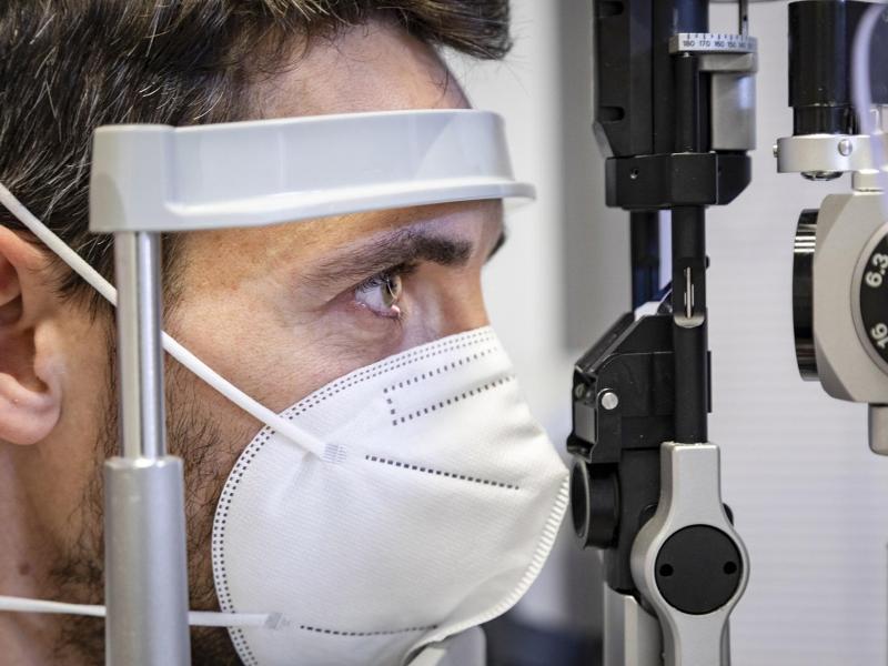
Diagnosing ocular nystagmus involves a comprehensive medical evaluation that combines patient history, eye examination, and specialized tests. Doctors first assess when symptoms began, their pattern, and any associated signs such as dizziness, neurological issues, or family history.
A detailed ophthalmological exam is then performed to evaluate visual acuity, eye structures, and eye movements, sometimes supported by tests like ERG or VEP.
In many cases, brain imaging (especially MRI) is used to detect neurological causes, while vestibular tests help assess inner ear function. Additional tools such as OCT or genetic testing may be used when a structural or hereditary cause is suspected.
Treatment options for ocular nystagmus
Treatment for nystagmus is highly individualized and fundamentally depends on the underlying cause. The primary goal is to improve visual function, reduce oscillopsia, and address the root pathology when possible.
Treating the underlying cause
- BPPV: The Epley maneuver and other canalith repositioning procedures are effective in resolving vestibular nystagmus in up to 80% of BPPV cases.
- Infections/Inflammation: Antibiotic or antiviral therapy (e.g., for labyrinthitis) combined with anti-inflammatory agents (corticosteroids) can reduce or eliminate nystagmus.
- MS-related nystagmus: Disease-modifying therapies (DMTs) can reduce relapse frequency; symptomatic management is also important.
- Drug-induced nystagmus: Reducing or stopping the offending medication typically resolves the condition.
- Nutritional deficiency (Wernicke's): Intravenous thiamine (Vitamin B1) administration can reverse nystagmus if initiated promptly.
Optical and refractive correction
Correcting underlying refractive errors with glasses or contact lenses does not cure nystagmus but can maximize residual visual acuity. Contact lenses may be preferred over glasses in some patients, as they reduce the magnification effect and remain centered with eye movements.
Pharmacological treatments
Several medications have been studied for symptomatic control of nystagmus, particularly oscillopsia:
- Memantine (an NMDA receptor antagonist): Shown to reduce nystagmus intensity in a randomized controlled trial
- Gabapentin: Demonstrated efficacy in acquired pendular nystagmus and downbeat nystagmus
- Baclofen: Effective in periodic alternating nystagmus, reducing the intensity of oscillations
- 4-Aminopyridine (4-AP): A potassium channel blocker showing promising results in downbeat nystagmus associated with cerebellar atrophy
- Clonazepam: Occasionally used in vestibular forms
- Acetazolamide: Used in specific hereditary forms (e.g., episodic ataxia with nystagmus)
Note: The use of these medications should always be supervised by a neurologist or ophthalmologist, as none are universally effective and all carry potential side effects.
Prism lenses
Optical prisms can shift the visual field to align with the null point, reducing the need for compensatory head posture and potentially improving visual comfort.
Botulinum toxin (Botox)
Injection of botulinum toxin into the extraocular muscles has been used in refractory cases to temporarily reduce the amplitude of nystagmus. While not a long-term solution, it can offer significant symptomatic relief, particularly for acquired pendular nystagmus with disabling oscillopsia.
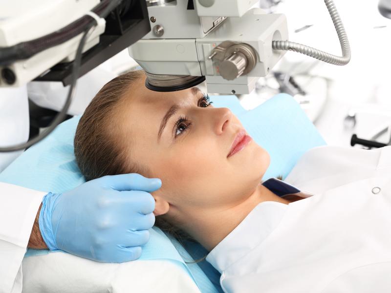
Surgical interventions
Surgery is not curative for most forms of nystagmus but may be considered in specific situations:
- Kestenbaum-Anderson procedure: Is eye muscle surgery that shifts the null point toward central gaze, reducing head turn and often improving visual function. Studies generally report functional improvement in many patients, with variable outcomes depending on the case.
- Refractive surgery (LASIK/PRK): Can correct associated refractive errors but does not directly treat nystagmus.
- Cataract surgery: In congenital nystagmus associated with dense cataracts, early surgical removal and visual rehabilitation can dramatically improve outcomes if performed in the first weeks of life.
Low vision rehabilitation
For patients with persistent visual impairment despite treatment, low vision rehabilitation is an essential component of care:
- Magnifying lenses and electronic magnifiers
- Tinted lenses (particularly beneficial in albinism-related nystagmus)
- Occupational therapy for daily living activities
- Educational support for children
Nystagmus in children: What parents should know
Nystagmus in infants and young children is particularly important to identify early. When a baby's eyes are observed to oscillate rhythmically and persistently after 3 months of age, a pediatric ophthalmologist should be consulted without delay.
Early intervention, whether optical, surgical (e.g., cataract removal), or rehabilitative, can significantly improve developmental visual outcomes. The brain's plasticity in early childhood means that timely treatment can maximize the potential for functional vision.
Children with nystagmus may also benefit from support in the school environment, including preferential front-row seating, enlarged print materials, and additional time for visual tasks.

"Medical journalist specializing in science communication, I put my expertise at the service of clear and accessible information. For Turquie Santé, I create content based on up-to-date medical data, in collaboration with specialists from partner clinics. My commitment is to provide reliable, transparent information that complies with international medical standards."
Need a personalized medical opinion?
Our partner doctors reply online within 24h, free of charge.