

Summary
- What is normal Intraocular Pressure (IOP)?
- What causes high eye pressure (Ocular hypertension)?
- Symptoms of ocular hypertension: The silent condition
- How is ocular hypertension diagnosed?
- What are the consequences of untreated ocular hypertension?
- Treatment options for ocular hypertension in Turkey
- Prevention and monitoring: Protecting your vision long-term
- Treatment of ocular hypertension in Turkey
Ocular hypertension is defined as an increase in intraocular pressure above 21 mmHg, without documented damage to the optic nerve or loss of the visual field. This condition is considered one of the main risk factors for glaucoma, a disease that can lead to permanent blindness if left without appropriate medical care.
In most cases, patients do not experience any noticeable symptoms in the early stages, making regular eye examinations the most important preventive measure. In this medical guide, we explain everything you need to know: normal eye pressure values, possible causes, how the condition is diagnosed, and the available treatment options, including specialized care in Turkey.
What is normal Intraocular Pressure (IOP)?
Intraocular pressure (IOP) refers to the fluid pressure inside the eye. It is measured in millimetres of mercury (mmHg) and plays a critical role in maintaining the structural integrity of the eyeball and optimal light refraction for clear vision.
Reference ranges at a glance
| IOP Category | Value (mmHg) |
| Low (Ocular Hypotony) | Below 12 mmHg |
| Normal | 12 – 21 mmHg |
| Ocular Hypertension | 22 mmHg or above |
| Acute Glaucoma (emergency) | Above 30 mmHg |
Maintaining pressure within the 12–21 mmHg physiological range is essential for proper anatomical conditions for refraction and good visual acuity. When pressure is too low (ocular hypotony, below 12 mmHg), fluid may be leaking from the eyeball, potentially causing vision distortion or even structural collapse of the globe. When pressure exceeds 21–22 mmHg consistently, the eye is classified as hypertensive.
Important: IOP naturally fluctuates throughout the day, typically peaking in the morning and dropping in the evening. It also tends to increase with age: from the age of 40 onwards, IOP rises by approximately 1 mmHg per decade. This age-related increase makes regular eye check-ups especially important for adults over 40.
Statistic: Studies estimate that ocular hypertension affects approximately 3–6% of adults, with prevalence increasing with age. According to the Ocular Hypertension Treatment Study (OHTS), around 9–10% of untreated patients with elevated intraocular pressure and additional risk factors may develop glaucoma over a 5-year period. However, individual risk varies significantly depending on clinical characteristics such as corneal thickness, baseline intraocular pressure, and optic nerve structure.
What causes high eye pressure (Ocular hypertension)?
Intraocular pressure is regulated by the production and drainage of aqueous humour, a clear fluid produced by the ciliary body that fills the anterior chamber of the eye (the space between the iris and the cornea). Any disruption to this delicate balance can raise pressure.
Primary mechanisms
- Overproduction of aqueous humour: The ciliary body produces fluid faster than it can drain.
- Impaired drainage: Blockage or dysfunction of the trabecular meshwork (the eye's drainage system) prevents normal fluid outflow.
- Combined mechanism: Both overproduction and reduced drainage occurring simultaneously.
Common risk factors and causes
- Medications, particularly corticosteroids (eye drops, oral, inhaled or injected)
- Eye trauma or injury affecting the drainage angle
- Ocular diseases such as pseudo-exfoliation syndrome, pigment dispersion syndrome, or uveitis
- Systemic conditions: diabetes mellitus, cardiovascular disease, arterial hypertension
- Chronic psychological stress and high cortisol levels
- High-risk pregnancies with sudden haemodynamic changes
- Smoking (associated with impaired ocular blood flow)
- A poor diet high in salt, refined sugar, alcohol, and processed red meat
- Genetic predisposition and family history of glaucoma
- Anatomical factors such as a naturally narrow drainage angle
Transient (Temporary) pressure spikes
Not all elevated IOP readings indicate chronic ocular hypertension. Pressure can rise transiently during:
- Vigorous physical exercise, especially heavy weightlifting
- Episodes of coughing, sneezing, or vomiting
- Consuming large amounts of caffeine or alcohol
- Lying in certain positions (IOP tends to be higher when lying flat)
A single elevated reading does not diagnose ocular hypertension. Diagnosis requires repeated measurements over multiple visits.
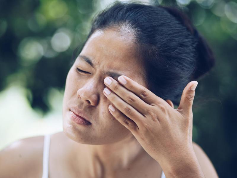
Symptoms of ocular hypertension: The silent condition
One of the most dangerous aspects of ocular hypertension is that, in the vast majority of cases, it causes no noticeable symptoms in the early stages. This is why it is sometimes called 'the silent thief of sight'.
Early stage: Usually asymptomatic
Most people with elevated intraocular pressure feel nothing unusual. There is no pain, no visual disturbance, and no external sign that something is wrong. This is precisely why routine eye examinations including IOP measurement are so important.
Possible non-specific signs
In some cases, patients may notice:
- Mild redness of the eyes
- Excessive tearing
- A sensation of eye fullness or pressure
- Occasional mild discomfort (though not sharp pain)
Late-stage symptoms (After optic nerve damage has begun)
After years of sustained elevated pressure, optic nerve damage can begin to develop. At this stage, patients may experience:
- Progressive peripheral (side) vision loss, often the first glaucoma symptom
- A persistent 'foggy' or 'misty' sensation in the visual field
- Increasing difficulty focusing, especially in low light
- In acute angle-closure glaucoma: sudden severe eye pain, nausea, rainbow halos around lights, and rapid vision loss
When to seek emergency care: If you experience sudden, intense eye pain, sudden vision loss, nausea with eye pain, or see coloured halos around lights, go to an ophthalmology emergency unit immediately. These may indicate an acute glaucoma attack, which can cause permanent vision loss within hours.
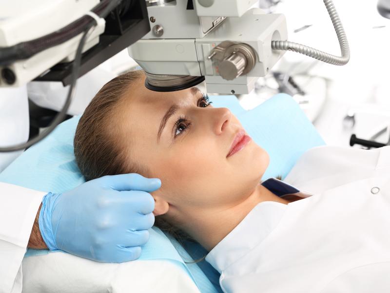
How is ocular hypertension diagnosed?
Because the condition is often asymptomatic, diagnosis depends entirely on a professional eye examination. The following tests are typically used:
Tonometry
The primary test for measuring IOP. The most common methods are:
- Goldmann Applanation Tonometry (GAT): Considered the gold standard, a small probe gently touches the cornea to measure pressure.
- Non-contact tonometry (air puff test): A brief puff of air is directed at the eye, no contact required, useful for screening.
- Rebound tonometry (e.g., iCare): A tiny probe briefly touches the cornea; often used in children or uncooperative patients.
Additional examinations
- Ophthalmoscopy / Fundus examination: The ophthalmologist examines the optic disc for signs of cupping or damage.
- Pachymetry: Measures corneal thickness, which affects IOP readings. Thin corneas may underestimate true IOP.
- Visual field testing (perimetry): Maps peripheral vision to detect early glaucomatous defects.
- Gonioscopy: Examines the drainage angle to classify the type of glaucoma risk.
- OCT (Optical Coherence Tomography): Provides a detailed scan of the optic nerve fibre layer to detect structural damage before symptoms appear.
Diagnosis typically requires several measurements over time, as IOP fluctuates. A single elevated reading is not sufficient to confirm ocular hypertension.
What are the consequences of untreated ocular hypertension?
Persistently elevated intraocular pressure is the primary risk factor for open-angle glaucoma, the most common form of glaucoma. Unlike acute-angle closure glaucoma, open-angle glaucoma develops slowly and painlessly. The optic nerve fibres gradually degrade, and peripheral vision is lost progressively, often without the patient noticing until significant damage has occurred.
Glaucoma is irreversible: once optic nerve fibres are destroyed, they cannot regenerate. This makes prevention and early treatment absolutely critical.
Key fact: Not everyone with ocular hypertension develops glaucoma, and conversely, glaucoma can occur in people with normal IOP (so-called normal-tension glaucoma). This is why a comprehensive eye examination, not IOP measurement alone, is essential for risk stratification.
Treatment options for ocular hypertension in Turkey
The goal of treatment is to lower intraocular pressure to a level that reduces the risk of optic nerve damage. The appropriate approach depends on the level of IOP, the presence of other risk factors, and the patient's overall health profile.
Medical treatment (First-line)
- Prostaglandin analogues (e.g., latanoprost, bimatoprost): Increase aqueous drainage. Once-daily dosing. Considered the most effective class.
- Beta-blockers (e.g., timolol): Reduce aqueous production. Often combined with other agents.
- Carbonic anhydrase inhibitors (e.g., dorzolamide, brinzolamide): Reduce fluid production.
- Alpha-2 agonists (e.g., brimonidine): Reduce production and increase drainage.
- Rho kinase inhibitors (e.g., netarsudil): Newer class improving trabecular outflow.
Laser treatment
- Selective Laser Trabeculoplasty (SLT): A minimally invasive laser procedure that improves trabecular drainage. Often effective as a first-line or adjunct treatment, with a good safety profile.
- Laser Peripheral Iridotomy (LPI): Used in narrow-angle or angle-closure cases.
Surgical options
- Trabeculectomy: Creates a new drainage channel. Reserved for advanced or refractory cases.
- MIGS (Minimally Invasive Glaucoma Surgery): Newer techniques such as trabecular micro-bypass stents with a faster recovery profile.
Lifestyle measures (Adjunctive)
While not a replacement for medical treatment, the following lifestyle adjustments may support IOP management:
- Regular moderate aerobic exercise (e.g., walking, swimming) has been shown to modestly reduce IOP
- Reducing caffeine and alcohol consumption
- Maintaining a healthy body weight and blood pressure
- A diet rich in leafy greens, antioxidants (vitamins C, E), and omega-3 fatty acids
- Sleeping with the head slightly elevated (10–20°)
- Stress management through mindfulness, yoga, or relaxation techniques
- Avoiding tight collars or neckties that can transiently increase IOP
Prevention and monitoring: Protecting your vision long-term
While ocular hypertension cannot always be prevented, its consequences can be minimised through early detection and consistent monitoring.
Recommended eye examination frequency
- Adults under 40 with no risk factors: Every 5 to 10 years
- Adults 40–54: Every 2 to 4 years
- Adults 55–64: Every 1 to 3 years
- Adults 65 and older: Every 1 to 2 years
- High-risk individuals (family history, diabetes, hypertension): Annually or as directed
Who should be screened?
You should consider an ophthalmological evaluation including IOP measurement if you:
- Are over 40 years of age
- Have a first-degree relative with glaucoma
- Have diabetes, cardiovascular disease, or arterial hypertension
- Are of African or Hispanic descent (higher genetic risk for glaucoma)
- Use corticosteroid medications regularly (inhaled, topical, or systemic)
- Have previously suffered an eye injury
Treatment of ocular hypertension in Turkey
In Turkey, ocular hypertension is managed according to internationally accepted ophthalmology guidelines, with the main goal of lowering intraocular pressure and preventing progression to glaucoma.
Treatment usually starts with medicated eye drops, such as prostaglandin analogs or beta-blockers, which help reduce eye pressure and are commonly used as first-line therapy. Patients are closely monitored to assess response and adjust treatment if needed.
If eye drops are not sufficient or not well tolerated, laser treatment (most commonly selective laser trabeculoplasty) may be recommended to improve fluid drainage and lower intraocular pressure.
In more complex or resistant cases, surgical options can be considered, although this is less common for ocular hypertension alone.
Ophthalmology centers in Turkey typically provide full diagnostic evaluation and long-term follow-up using modern imaging techniques, ensuring early detection of any progression toward glaucoma.

"Medical journalist specializing in science communication, I put my expertise at the service of clear and accessible information. For Turquie Santé, I create content based on up-to-date medical data, in collaboration with specialists from partner clinics. My commitment is to provide reliable, transparent information that complies with international medical standards."
Need a personalized medical opinion?
Our partner doctors reply online within 24h, free of charge.