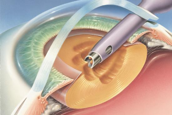
Retinopathy of prematurity is a common problem in preterm infants. It is the cause of most eye problems in former premature babies even after treatment. But more importantly, it is one of the leading causes of blindness in children.
The disease can be treated if it is diagnosed in time. This is why pediatric doctors are working to evolve the treatments for this disease to guarantee these newborns a normal life.
Any Question ?
Get in Touch.
We're Here to Help.

What is retinopathy of prematurity?
Retinopathy of prematurity (ROP),also called retrolental fibroplasia, is a disorder of the developing retinal blood vessels in premature infants.
The disease develops when the blood vessels grow unusually. It leads to the formation of a knot of tissue between the central retina and the peripheral retina. The risk of retinopathy of prematurity is closely related to the proportion of retinal surface area not vascularized at birth.
The chances of a newborn baby weighing less than 1 kg developing retinopathy are very high. Indeed, the disease occurs in 47 to 80% of cases where the infant weighs less than 1 kg. It can also increase in case of repeated clinical complications. A severe form of the disease manifests itself in 21-43% of these cases.
The different stages of retrolental fibroplasia
The classification of retinopathy of prematurity is done according to the stages of the disease as in the following table:
Stage 1 | Demarcation line. |
Stage 2 | Knot of tissue forming in the dividing line. |
Stage 3 | Extra-retinal fibrovascular multiplication. |
Stage 4 | Partial retinal detachment. |
Stage 5 | Total retinal detachment. |
What causes retinopathy of prematurity?
Retinopathy of prematurity depends on many causes and factors that increase the risk of its development. These disease-promoting factors are multiple, but there are two main ones:
Oxygen-dependent factors
Historically, the first risk factor for retinopathy that has been identified is poorly controlled oxygen therapy. Indeed, hyper-oxygenation of the retina of a newborn baby can cause retinopathy in premature babies. It causes damage caused by increased oxygen levels in addition to reduced levels of vascular growth factors.
Oxygen independent factors
In addition to oxygen therapy, other major risk factors for retinopathy of prematurity have been identified. These factors are:
- Low gestational age.
- Low birth weight.
- The quality of postnatal growth.
When and how should retinopathy of prematurity be diagnosed?
The retinopathy of prematurity develops between the 32nd and 34th weeks of gestation. So, the screening for the disease is usually done from the 31st week of gestation at birth. It is also performed in all newborns weighing less than 1.5 kg or who were born before 30 weeks of gestation.
The pediatricians diagnose the retinopathy of prematurity is by a fundoscopic examination, also called an ophthalmoscopy. If the disease is detected, the examination may reveal two results:
- Benign case: A line of demarcation and a ridge.
- More serious case: Multiplication of the blood vessels of the retina.
Treatment of retinopathy of prematurity in Istanbul's clinics
To fight retinopathy of prematurity, specialist ophthalmologyclinics in Istanbul offer two major means of treatment:
The laser photocoagulation
Laser photocoagulation is a treatment used for severe retinopathy of prematurity. It is focused on the avascular part and it allows the peripheral retina to be removed. This treatment aims to reduce the incidence of retinal detachment, as well as the production of blood vessel growth factors.
It is necessary to monitor the blood supply to the retina every 1 to 2 weeks until the blood vessels mature.
There are similar procedures, but which remain of last resort because they bring little benefit. One of these procedures is vitrectomy with lens removal for retinal detachment in childhood.
A newborn baby who suffers from complete retinal detachment should be monitored to diagnose the emergence of secondary glaucoma.
If the laser photocoagulation patient has residual scarring (scar tissue),he will need lifelong follow-up at least once a year.
Treatment with Bevacizumab
Bevacizumab is an anti-vascular endothelial growth factor monoclonal antibody. Treatment with this anti-growth factor is a recent advance in the fight against retinopathy of prematurity. It helps stop the progression of the disease in newborns.
Compared with laser therapy, Bevacizumab shows a lower recurrence rate and a lower myopia prevalence.
Unfortunately, anti-factor therapy remains a second-line therapy despite its advantages. This is due to the accompanying drawbacks, including:
- Need for large dose of Bévacizumab.
- Risk of infection.
There are other treatments for retinopathy of prematurity such as scleral buckle.
Treating retinopathy of prematurity in Turkey: at which cost?
The clinics in Turkey with which we are partners offer the appropriate treatment for the good health of your children. In addition, they are internationally renowned. Indeed, they offer the services of internationally recognized doctors. Added to this, a professional and attentive hospital staff who will make your stay easier.
Finally, our partner clinics offer the best possible rates. The total cost of treatment varies depending on the hospital and their location. However, it is still lower than that offered by other European clinics while maintaining the same quality of therapy.
You can get a personalized quote and the price of the operation in the link below.
Share this page